
Define the biology
Choose the cells, material context, spatial relationship and function relevant to the question.
Bioprinting fundamentals
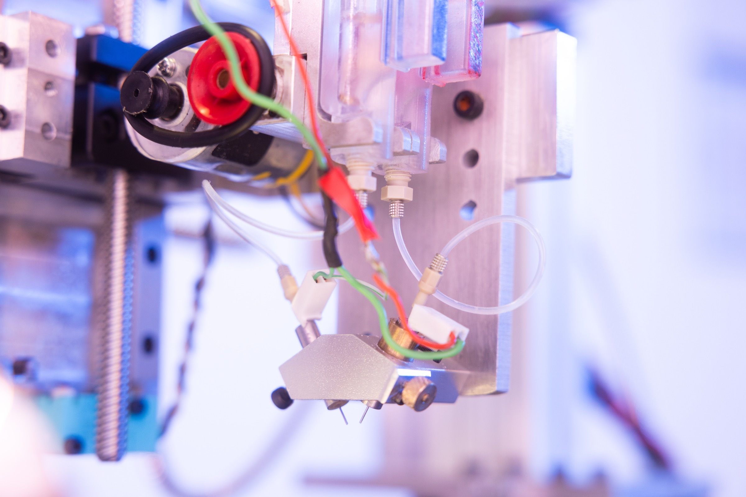
3D bioprinting uses controlled deposition to organise cells, biomaterials and other biological components into experimental structures. The value is not the shape alone. It is the ability to create a model that helps answer a defined biological question.
The basic idea
Cells in the body experience three-dimensional surroundings, neighbouring populations, extracellular matrix and mechanical cues. A bioprinted model can organise selected parts of that environment in a controlled laboratory format.
Choose the cells, material context, spatial relationship and function relevant to the question.
Match deposition and crosslinking to the material, resolution, cell-handling and geometry requirements.
Measure print quality, cell response, repeatability and the endpoint needed for scientific interpretation.
A model-development workflow
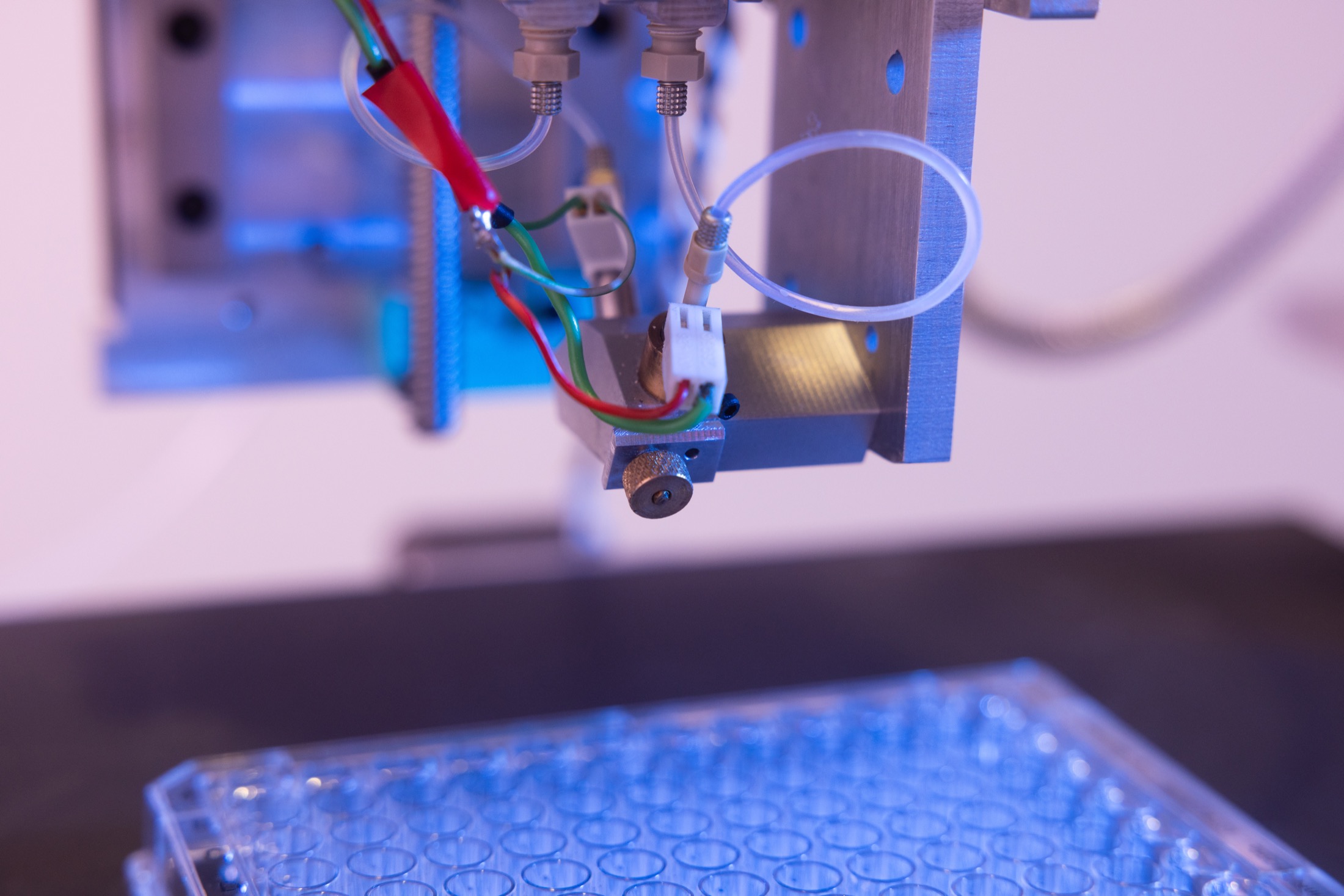
A useful workflow includes cell preparation, material formulation, deposition, crosslinking, culture, quality control and downstream analysis. Plate format, sterility, imaging and sampling can be as important as the print step.
When it may add value
JetBio's approach
JetBio combines ReJI, microvalve and inkjet development capabilities so teams can assess different deposition behaviours around the same biological brief. ReJI was first reported in peer-reviewed work on high-cell-density gels for bone microtissue fabrication.
Read the foundational ReJI publicationFrequently asked questions
3D printing creates structures from deposited materials. 3D bioprinting applies controlled deposition to biological materials and may include living cells, biomaterials and bioactive components, so cell handling and biological function become part of the process design.
Usually not. Most research models reproduce selected structures, cell interactions or functions needed to study a defined question. Their usefulness depends on the intended context and the evidence supporting that use.
No. Printability depends on material properties, crosslinking, cell requirements, deposition method, geometry and handling conditions. Compatibility should be assessed rather than assumed.
It is worth considering when manual culture or another fabrication method cannot reliably create the required cell density, spatial pattern, multi-material structure, substrate interface or repeatable format.
Start a useful conversation
Share the cells, materials, format and endpoint. JetBio can help assess whether bioprinting is a credible route.